Catalogue
-
-
- Products
-
- Mouse anti PSMA

Mouse anti PSMA
Catalog number: MUB1510P| Clone | 107.1A4 |
| Isotype | IgG1 |
| Product Type |
Monoclonal Antibody Primary Antibodies |
| Units | 0.1 mg |
| Host | Mouse |
| Species Reactivity |
Human |
| Application |
ELISA Flow Cytometry Immunocytochemistry Immunohistochemistry (frozen) Immunohistochemistry (paraffin) |
Background
Prostate specific antigen (PSA) is the most widely used marker of prostate cancer. Immunoassays for PSA using monoclonal (MAb) or polyclonal antibodies have clinical applications, such as monitoring and early detection of prostate cancer. However, PSA is not a perfect tumor marker because Serum levels often are elevated in men with benign prostatic hyperplasia, prostatitis and other nonmalignant disorders and also because levels are not always elevated in individuals with early prostate cancer. 107-1A4 is a new prostate cancer-reactive MAb which can identify additional proteins that could be of value for the diagnosis and treatment of prostate cancer. It shows selective reactivity to prostate epithelial membrane antigens.
Source
107.1A4 is a Mouse monoclonal IgG1/k antibody derived by fusion of SP2/0-Ag14 Mouse myeloma cells with spleen cells from an immunized Mouse, using a two-phase immunization protocol involving the prostate cancer cell line LNCaP.
Product
Each vial contains 100 ul 1 mg/ml purified monoclonal antibody in PBS containing 0.09% sodium azide.
Formulation: Each vial contains 100 ul 1 mg/ml purified monoclonal antibody in PBS containing 0.09% sodium azide.
Specificity
107.1A4 recognizes an antigen, which appeared to be distinct from those previously described and shows specific tumor targeting in preliminary in vivo studies. MAb 107-1A4 is suggested to recognize a distinct conformational and unique epitope in the extracellular domain of PSMA. PSMA is a type II membrane glycoprotein of 100kDa that has a short intracellular N-terminal domain (residues 1 to 18), a transmembrane region (residues 19 to 43) and a large extracellular domain consisting of residues 44 to 750.
Applications
MAb 107-1A4 could not be used in Western blot, is useful for immunohistochemistry on frozen and paraffin-embedded tissues, immunocytochemistry, flow cytometry and ELISA. Optimal antibody dilution should be determined by titration; recommended range is 1:100 – 1:1000 for immunohistochemistry with avidin-biotinylated Horseradish peroxidase complex (ABC) as detection reagent.
Storage
The antibody is shipped at ambient temperature and may be stored at +4°C. For prolonged storage prepare appropriate aliquots and store at or below -20°C. Prior to use, an aliquot is thawed slowly in the dark at ambient temperature, spun down again and used to prepare working dilutions by adding sterile phosphate buffered saline (PBS, pH 7.2). Repeated thawing and freezing should be avoided. Working dilutions should be stored at +4°C, not refrozen, and preferably used the same day. If a slight precipitation occurs upon storage, this should be removed by centrifugation. It will not affect the performance or the concentration of the product.
Caution
This product is intended FOR RESEARCH USE ONLY, and FOR TESTS IN VITRO, not for use in diagnostic or therapeutic procedures involving humans or animals. It may contain hazardous ingredients. Please refer to the Safety Data Sheets (SDS) for additional information and proper handling procedures. Dispose product remainders according to local regulations.This datasheet is as accurate as reasonably achievable, but our company accepts no liability for any inaccuracies or omissions in this information.
References
1. Brown, L.G., Wegner, S.K., Wang, H., Buhler, K.R., Arfman, E.W., Lange, P.H. and Vesella,. (1998). A novel monoclonal antibody 107-1A4 with high prostate specificity: generation, characterization of antigen expression, and targeting of human prostate cancer xenografts. Prostate Cancer Prostatic Dis. 1, 208-15.
2. Wang, S., Diamond, D.L., Hass, G.M., Sokoloff, R. and Vesella, R.L. (2001). IdentifiCation of prostate specific membrane antigen (PSMA) as the target of monoclonal antibody 107-1A4 by proteinchip; array, surface-enhanced laser desorption/ionization (SELDI) technology. Int J Cancer 92, 871-6.
Safety Datasheet(s) for this product:
| NM_Sodium Azide |

|
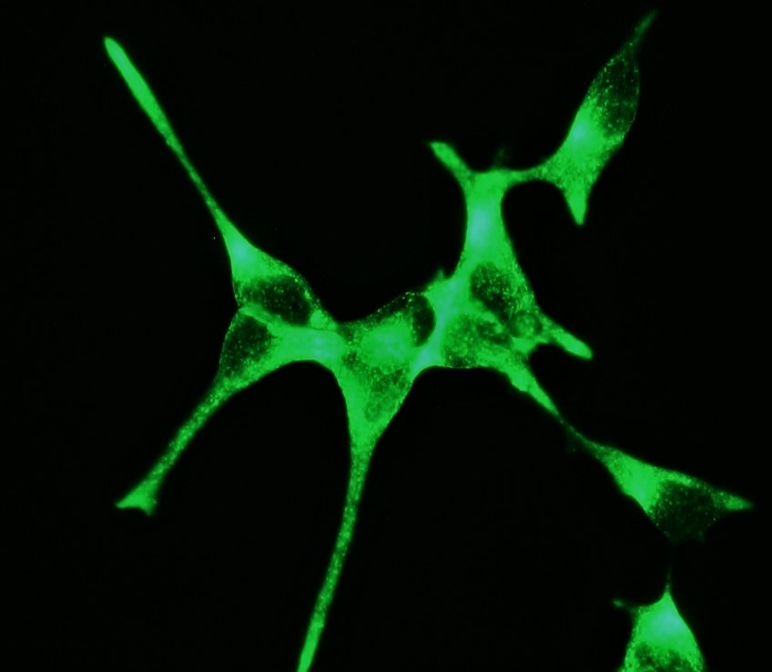
Figure 1. Indirect immunofluorescence staining of PSMA in the prostate cancer cell line LNCaP cells using MUB1510P, clone 107-1A4. Note the membranous localization of PSMA. |
|
Figure 2. Indirect immunofluorescence staining of PSMA in the prostate cancer cell line LNCaP cells using MUB1510P, clone 107-1A4 (diluted 1:500). Note the membranous localization of PSMA. |
|
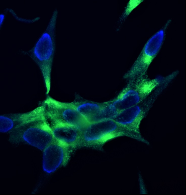
Figure 3. Indirect immunofluorescence staining of PSMA in the prostate cancer cell line LNCaP cells using MUB1510P, clone 107-1A4 (diluted 1:500). Note the membranous localization of PSMA. Nuclear DNA staining with DAPI. |
|
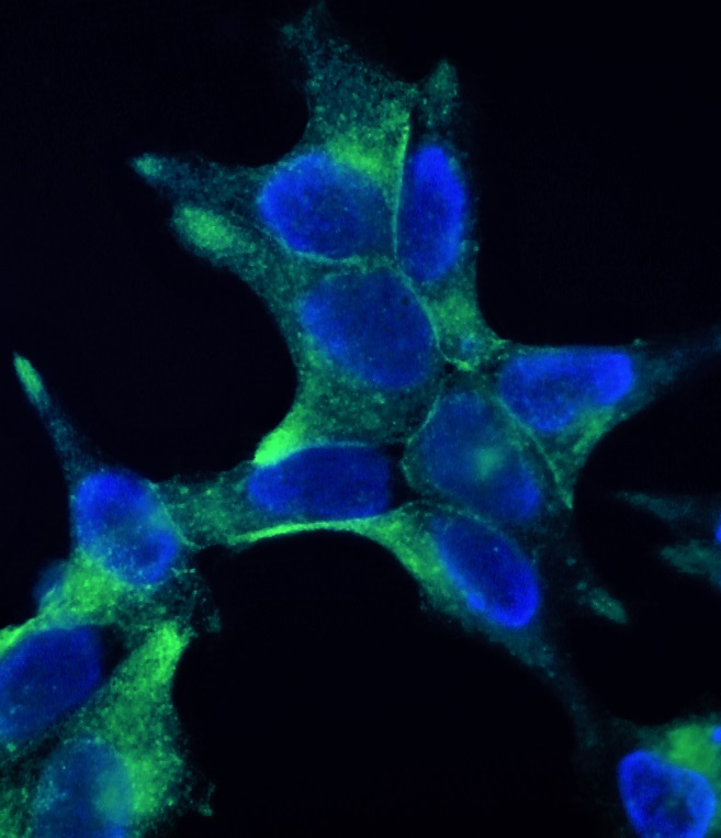
Figure 4. Indirect immunofluorescence staining of PSMA in the prostate cancer cell line LNCaP cells using MUB1510P, clone 107-1A4 (diluted 1:500). Note the membranous localization of PSMA. Nuclear DNA staining with DAPI. |

Figure 1. Indirect immunofluorescence staining of PSMA in the prostate cancer cell line LNCaP cells using MUB1510P, clone 107-1A4. Note the membranous localization of PSMA.

Figure 2. Indirect immunofluorescence staining of PSMA in the prostate cancer cell line LNCaP cells using MUB1510P, clone 107-1A4 (diluted 1:500). Note the membranous localization of PSMA.

Figure 3. Indirect immunofluorescence staining of PSMA in the prostate cancer cell line LNCaP cells using MUB1510P, clone 107-1A4 (diluted 1:500). Note the membranous localization of PSMA. Nuclear DNA staining with DAPI.

Figure 4. Indirect immunofluorescence staining of PSMA in the prostate cancer cell line LNCaP cells using MUB1510P, clone 107-1A4 (diluted 1:500). Note the membranous localization of PSMA. Nuclear DNA staining with DAPI.