Catalogue

SCICONS anti-dsRNA antibody (K1)
Catalog number: 10020500| Clone | K1 |
| Isotype | IgG2a kappa |
| Product Type |
Monoclonal Antibody |
| Units | 500 µg |
| Host | Mouse |
| Application |
ELISA Flow Cytometry Immuno-affinity-chromatography Immunoblotting Immunocytochemistry Immunohistochemistry |
Background
This product is part of the SCICONS™ product line which offers the gold standard in anti-dsRNA antibodies.
Over the past decade our double-stranded RNA (dsRNA)antibodies have been used extensively to detect and characterise plant and animal viruses with dsRNA genomes or intermediates. In addition, the anti-dsRNA antibodies can be used as a diagnostic tool to detect pathogens, including detection in paraffin-embedded fixed tissue samples (Richardson et al. 2010).
The K1 monoclonal antibody recognises dsRNA with similar affinity to our widely used J2 antibody. It can be used for the histological and cytological detection of dsRNA in cells and tissues.
It has proven especially useful as an alternative to J2 to resolve cross-reactions and/or remove unwanted background, in those rare experimental setups where J2 did not provide satisfactory results.
K1 can be used to detect dsRNA intermediates of viruses as diverse as Hepatitis virus, Theiler’s murine encephalomyelitis virus or Japanese encephalitis virus. It has been for the detection of dsRNA in cultured cells and in fixed paraffin-embedded histological samples (see publications).
If Poly I:C needs to be detected we highly using K1 rather than J2 because K1 has a much higher affinity for this synthetic polyribonucleotide (see Schönborn et al. 1991, Fig. 2).
K1 has been used successfully in immunofluorescence microscopy, in flow cytometry (FACS) and in immunocapture methods (such as dot-blot and ELISA).
Synonyms: Mouse anti dsRNA
Source
Female DBA/2 mice were injected intraperitonially with a mixture of 50 ug L-dsRNA and 75 ug methylated bovine serum albumin, emulsified in complete Freund's adjuvant. After several boosts spleen cells were fused with Sp2/0-Agl4 myeloma cells to generate the hybridoma clone.
Product
The mAb K1 recognises double-stranded RNA (dsRNA) provided that the length of the helix is greater than or equal to 40 bp. dsRNArecognition is independent of the sequence and nucleotide composition of the antigen. All naturally occurring dsRNAs investigated up to now (40-50 species) as well as poly(I).poly(C) and poly(A).poly(U) have been recognised by K1. As described by Schönborn et al. K1 shows higher affinity to poly(I).poly(C) than to the other dsRNA antigens, although the difference of apparent binding constants may vary under different experimental conditions.
Formulation: The lyophilised sample should be reconstituted with 500 µl sterile distilled water. The mAb will then be in PBS without any stabilisers or preservatives at a concentration of 1 mgr/ml. As a result of the lyophilisation procedure, the reconstituted antibody may contain small amounts of denatured protein in the form of aggregates that may interfere with some applications such as immunohistochemistry (e.g. by giving high backgrounds). We therefore highly recommend centrifuging (microcentrifuge) the reconstituted antibody before use and using the supernatant.
Purification Method: Affinity chromatography on Protein A-agarose.
Purity: Gel electrophoretically pure IgG antibody.
Concentration: Concentration after reconstitution: 1.00 mg/ml as determined by A280 nm (A280 nm = 1.47 corresponds to 1 mg/ml antibody).
Applications
MAb K1 can be used for ELISA, dsRNA-immunoblotting, immuno-affinity-chromatography and in certain systems also for immunohistochemistry (see references).
The optimum working dilution of the antibody for any specific application should be established by titration.
Please note that nucleic acid separation prior to dsRNA-immunoblotting must be carried out by polyacrylamide gel electrophoresis, because the sensitivity of detection is considerably lower after blotting from agarose gels.
Not for use for clinical purposes. For in vitro use only.
Storage
After reconstitution antibodies should be aliquoted and stored at -20 °C or -70 °C. After adding 10 mM sodium azide undiluted antibody can also be stored at +4 °C for a short period of time. For long term storage the mAb should be kept frozen. Repeated freezing/thawing cycles should be avoided. When kept lyophilized the product will remain stable for 10 years at -20 °C or -70°C.
Shipping Conditions: Ship at ambient temperature.
Caution
This product is intended FOR RESEARCH USE ONLY, and FOR TESTS IN VITRO, not for use in diagnostic or therapeutic procedures involving humans or animals. It may contain hazardous ingredients. Please refer to the Safety Data Sheets (SDS) for additional information and proper handling procedures. Dispose product remainders according to local regulations.This datasheet is as accurate as reasonably achievable, but Nordic-MUbio accepts no liability for any inaccuracies or omissions in this information.
References
1) Schönborn, J., Oberstrass, J., Breyel, E., Tittgen, J., Schumacher, J. and Lukacs, N. (1991) Monoclonal antibodies to double-stranded RNA as probes of RNA structure in crude nucleic acid extracts. Nucleic Acids Res.19, 2993-3000.
2) Lukacs, N. (1994) Detection of virus infection in plants and differentiation between coexisting viruses by monoclonal antibodies to double-stranded RNA. J. Virol. Methods 47, 255-272.
3) Lukacs, N. (1997) Detection of sense:antisense duplexes by structurespecific anti-RNA antibodies. In: Antisense Technology. A Practical Approach, C. Lichtenstein and W. Nellen (eds), pp. 281-295. IRL Press, Oxford.
4) S. J. Richardson, A. Willcox, D. A. Hilton, S. Tauriainen, H. Hyoty, A. J. Bone, A. K. Foulis, N. G. Morgan. Use of antisera directed against dsRNA to detect viral infections in formalin-fixed paraffin-embedded tissue. J Clin Virol. (2010) 49(3); 180-5. doi: 10.1016/j.jcv.2010.07.015.
Safety Datasheet(s) for this product:
| SDS antibodies without sodium azide Nordic |

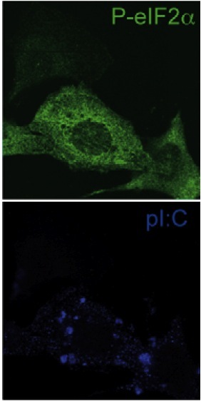
Figure 1. K1 antibody is used to detect dsRNA in MEFs treated with poly I:C (lower panel). Presence of dsRNA is concomitant with phosphorylation of eIF2a and thus protein synthesis arrest (upper panel). Figure taken from Clavarino et al.(2012) PLoS Pathog 8:e1002708.

